대장암의 위치에 따른 생존율 분석: 단일 술자 경험
Survival analysis for colon subsite and rectal cancers: Experience from a single surgeon
Article information
Trans Abstract
Purpose:
The survival rates of patients with colorectal cancers have been well documented in many studies. Some studies have shown that proximal colon cancers have inferior survival rates when compared with distal colon cancers. However, the prognostic significance of tumor location with respect to survival remains controversial. By using data from a single physician, we analysed patient survival rates based on colon cancer subsite location, including rectal cancers.
Methods:
We retrospectively analysed 881 patients with colorectal cancers between 1987 and 2008. Colon subsite locations were defined as cecum, ascending colon, transverse colon, descending colon, sigmoid colon, and rectum. Subsite-specific survival analyses were performed using Kaplan-Meier analysis and Cox proportional hazards ratios. The median follow-up time was 93 months.
Results:
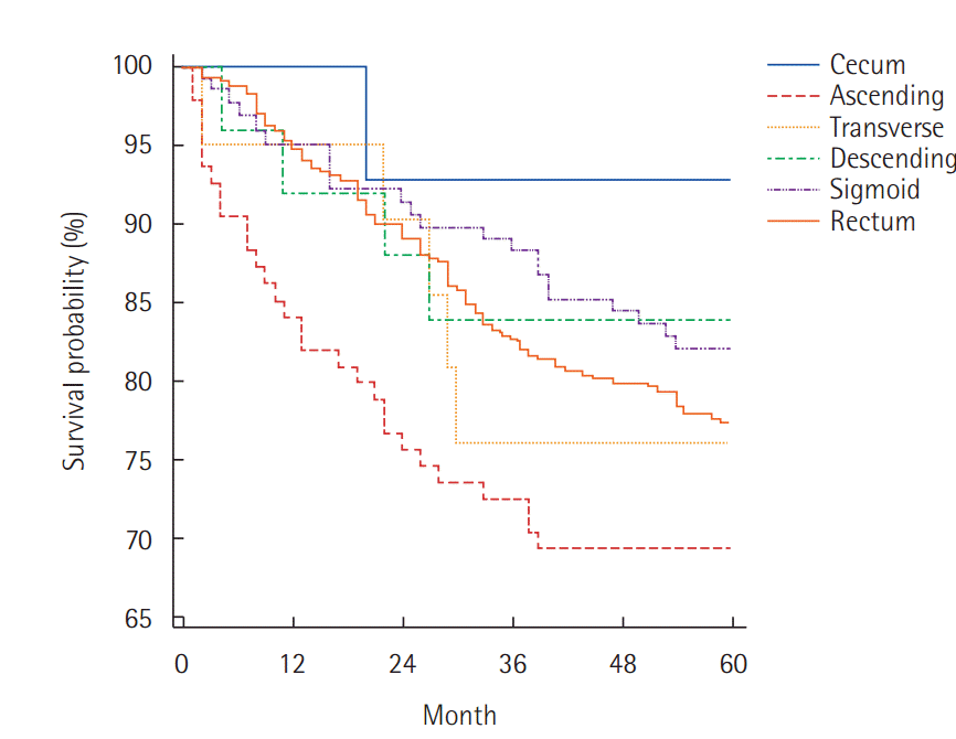
A total of 689 colorectal cancer cases were included in our analysis, of which 14 were cecum (2.0%), 95 were ascending colon (13.8%), 21 were transverse colon (3.0%), 25 were descending colon (3.6%), 129 were sigmoid colon (18.7%), and 405 were rectum (58.8%) cancers. The 5-year overall survival rates were 77.8% for all colorectal cancers, which consisted of 92.9% for cecal cancer, 69.5% for ascending colon cancer, 76.2% for transverse colon cancer, 84.0% for descending colon cancer, 82.2% for sigmoid colon cancer, and 77.5% for rectal cancer.
Conclusion:
Ascending colon cancer was associated with the poorest survival outcome, whereas descending colon cancer was associated with the best survival outcome except cecal cancer. Moreover, the survival rate associated with left colon cancer was better than the survival for right colon and rectal cancer.
INTRODUCTION
Colorectal cancer (CRC) is the third most common cancer in South Korea [1]. Survival rates associated with CRCs have been well documented in many studies. The proximal and distal colon and the rectum have different characteristics, including different embryologic origins, anatomical appearance and physiological functions. Additionally, colon and rectal cancers show differences in their biological features, treatment susceptibility profiles, and prognoses. Several studies have reported inconsistent results regarding the survival rates of colon cancer patients based on the location of the primary tumor. Some reports have shown superior survival in patients with left colon cancers after treatment than in patients with right colon cancers [2-5], whereas other studies reported no difference in the survival between these two patient cohorts [6,7]. Furthermore, survival comparisons between colon and rectal cancer patients have been inconsistent. Therefore, in this study, we compared the survival rates of patients with colon cancers at different subsites, including rectal cancers, using data accrued by a single surgeon.
METHODS
Study population
From January 1987 to December 2008, we retrospectively analysed 689 among 881 patients diagnosed with CRC and treated by a single surgeon. Patients who underwent palliative surgery, underwent surgery at another hospital, died within 30 days of surgery, or experienced hereditary colon cancers, such as familial adenomatous polyposis and hereditary nonpolyposis CRC, were excluded from this study. Variables for clinical characteristics included age, gender, anatomic tumor location, preoperative serum carcinoembryonic antigen (CEA) levels, tumor node and metastasis (TNM) stage, and lymph node ratio (LNR). Written informed consent was obtained from all patients before the operation. After Institutional Review Board approval of our hospital, all records and medical charts of patients were reviewed.
Classification of location
Subsite location of the tumors was defined as follows: cecum, ascending colon (includes ascending colon and hepatic flexure), transverse colon, descending colon (includes splenic flexure and descending colon), sigmoid colon (includes sigmoid colon and rectosigmoid junction), and rectum. Moreover, right or proximal colon included cancers of the cecum, ascending colon, hepatic flexure, and transverse colon. The left or distal colon included cancers of the splenic flexure, descending colon, sigmoid colon, and the rectosigmoid junction.
Statistical analysis
The chi-square test was used for comparing risk factor distributions between subsite groups. Overall survival rates were analysed using the Kaplan–Meier method, and statistical significance of the survival rates was evaluated using the log-rank test. Multivariate analysis was used to assess all of the clinicopathological factors associated with overall survival using Cox proportional hazards regression (i.e., age, sex, CRC subsite location, TNM stage, LNR, and preoperative serum CEA levels). A P-value of <0.05 was considered statistically significant. Statistical analysis was performed using MedCalc Statistical Software ver. 14.10.2 (MedCalc Software bvba, Ostend, Belgium).
RESULTS
Clinicopathological characteristics of CRC patients
A total of 689 CRC patients were included in the analysis with a median follow-up of 93 months (range, 1–193 months). Of these patient tumors, 14 tumors were located in the cecum, 95 in the ascending colon, 21 in the transverse colon, 25 in the descending colon, 129 in sigmoid colon, and 405 in the rectum. The TNM stage of the study population was I in 47 patients, II in 326 patients, III in 236 patients, and IV in 79 patients. The number of patients with tumors located at each subsite is listed in Table 1 according to patient sex, age, and TNM stage.
Overall survival of CRC patients
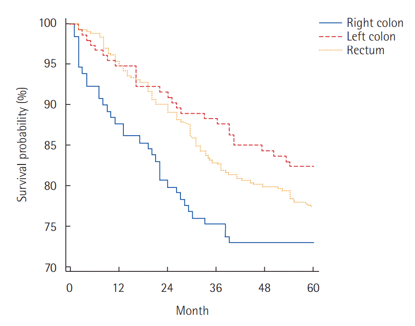
Overall survival rates according to cancer subsite location are shown in Table 2, and the overall survival curves according to tumor subsite location are presented in Fig. 1. The 5-year survival rate was 92.9% for patients with cancer of the cecum, 69.5% for patients with cancer of the ascending colon, 76.2% for patients with cancer of the transverse colon, 84.0% for patients with cancer of the descending colon, 82.2% for patients with cancer of the sigmoid colon, 77.5% for patients with cancer of the rectum, and 77.8% for all cancer patients combined. The overall survival curves after grouping the subsites according to right and left colon cancers are presented in Fig. 2. Right colon cancers were associated with a poorer survival rate (70.7%) than left colon cancers (82.5%) (P<0.05). There was no statistically significant difference in the 5-year survival rates between patients with colon cancer and rectal cancer (77.4% and 77.5%, respectively) (Table 3).
Survival analysis of stage I and II cancers showed no difference based on tumor location. However, stage III tumors showed statistically significant differences based on the location of the malignancy (P<0.05). Furthermore, stage III descending colon cancers were associated with a better survival rate than stage III tumors at any other location except cecum due to its rarity, whereas stage III rectal cancers were associated with the worst overall survival rate of all stage III tumor locations. In patients with stage IV tumors, no differences in the survival rates for any tumor location were observed. Additionally, right colon tumors were associated with worse survival when compared with left colon tumors (hazard ratio [HR], 0.31; 95% confidence interval [CI], 0.13–0.72) and rectal tumors (HR, 0.75; 95% CI, 0.35–1.61) (P<0.05) when analysing stage III tumors, but not when analysing stage I, II or IV tumors (Table 3). The variables associated with survival rate included serum CEA level, LNR, age over 65 years and CRC location. In the multivariate analysis, age, advanced CRC stage, serum CEA level, lymph node ratio and presence of sigmoid colon cancer or left colon cancer were associated with statistically significant survival differences (Table 4).
DISCUSSION
Due to their similar characteristics, colon and rectal cancers are often defined together as CRCs. Well-known prognostic factors for CRCs include age, sex, T stage, N stage, preoperative serum CEA level, tumor differentiation, lympho-vascular invasion, and perineural invasion [8]. Treatments for CRCs include surgical resection, chemotherapy and radiotherapy, of which adequate surgical resection is considered a cornerstone of CRC treatment. However, differences in CRC patient survival rates have been reported based on the accuracy of resection, the response of the tumor to chemotherapy, and the presence/absence of radiotherapy among other factors. Moreover, many studies have shown that colon cancers are different from rectal cancers in terms of their embryologic, environmental, genetic and biomolecular characteristics. In addition, right-sided colon cancers are associated with a different prognosis than left-sided colon and rectal cancers. We assumed that based on the tumor location, different CRCs may be treated differently in terms of surgery and chemoradiotherapy. Therefore, to reduce interpatient variability in treatment, our study was designed based on the data obtained from a single surgeon.
Studies have suggested that the anatomical site of the tumor is an important factor in the management of CRCs. However, site-specific CRC survival data remain controversial. Many studies have classified colon cancers as right or left, of which right colon cancers are associated with poorer prognosis [2-5,9-11]. For example, Elsaleh et al. [3] reported the 10-year survival rates to be 27% and 37% for right and left CRCs, respectively. Meguid et al. [2] demonstrated a 5% decrease in the survival rate for patients with right colon cancer when compared with patients with left colon cancers. In terms of disease-free survival, Park et al. [8] reported that the right colon cancer patients performed better than the left-side colon cancer patients. Moreover, Sjo et al. [12] showed improved survival in the patients with right colon cancer than in those with left colon cancers. Other studies involving rectal cancers showed that transverse colon cancers were associated with the worst outcome (HR, 1.25) [13]. Moreover, in another study, colon cancer patients showed superior survival than rectal cancer patients [14,15]. However, patients with advanced stage rectal cancer experienced better survival rates than advanced stage colon cancer patients [16].
In our study, proximal colon cancer patients experienced poorer survival rates than distal colon cancer patients. Ascending colon cancer was associated with the worst survival of all locations analysed. Moreover, descending colon cancer was associated with the best survival rate out of all cancer analysed except cecal cancer, which was excluded due to the small sample size. Nawa et al. [17] reported that right-sided cancers are more likely to be detected at an advanced stage. In our study, more patients with stage III tumors experienced left colon cancer, whereas more patients with stage IV tumors experienced right colon cancer (P<0.05). These results suggest that right-sided cancers tend to be less symptomatic in the earlier stages. Furthermore, surgical strategies in patients with left colon and rectal colon cancers include high ligation of the inferior mesenteric artery or the inferior mesenteric vein for access to the high-yield lymph nodes and inferior reach. In contrast, the strategy for lymph node harvest remains constant in patients with right-side colon cancers and consists of ligation of the mesocolic vessels near the superior mesenteric vessel. Moreover, complete mesocolic excision was not conducted in patients with advanced stage disease. Complete mesocolic excision is associated with improved outcomes for patients with colon cancers [18]. Additionally, the worst survival rate associated with ascending colon cancer patients is consistent with other studies [2,10] for reasons that remain unclear. Our results showing that the best survival rate was associated with descending colon cancers differs from other reports, which have shown that sigmoid colon cancers are associated with the best survival in our multivariate analysis and other study [2]. One possibility is that other studies misclassified some rectal cancers as rectosigmoid junction cancers to meet the conditions of national insurance coverage for adjuvant chemotherapy. This could result in a decrease in the survival rate of sigmoid colon cancer patients in our study. We observed no significant differences in the survival rates of colon cancer and rectal cancer patients, even in stage III patients. In addition, cecal cancers were associated with the best survival rates in our study; however, the sample size was too small for analysis. Majek et al. [19] previously reported survival results for cecal cancer patients and showed no survival advantage for patients with this type of tumor. The cecum is an independent colonic subsite that should be classified as a separate site; however, our study is insufficient to define a significant survival advantage in cecum cancer patients. We anticipate that a larger study of cecal cancer patients is necessary to show more accurate and reliable results.
Several limitations are associated with this study. Some cancer locations, including the cecum, the transverse colon and the descending colon, were represented by very few patients. Moreover, accurate grouping of junctional cancers, such as hepatic, splenic flexure and rectosigmoid cancers, could have influenced our results. Finally, biomolecular analysis of the tumors for the mutational and expression level statuses of genes such as KRAS, BRAF, p53, and microsatellite instability could not be obtained because a large proportion of the data were obtained too long before the analysis.
Among all CRCs, patients with ascending colon cancer showed the poorest survival outcome. Descending colon cancer was associated with the best overall survival in CRC patients to the exclusion of cecal cancer. Moreover, left colon cancers were associated with better survival than right colon cancers and rectal cancers. Therefore, our results suggest that more careful approaches are needed for CRC treatment depending on the localization of the tumor.
Notes
No potential conflict of interest relevant to this article was reported.
Acknowledgements
This study was supported by a clinical research grant from the Pusan National University Hospital in 2014.


