증상을 동반한 원발성 폐암의 위장관 전이 2예
Two cases of symptomatic gastrointestinal metastases from primary lung cancer
Article information
Trans Abstract
Primary lung cancer is relatively common malignant tumor throughout the world and often spreads to the contralateral lung, liver, brain, adrenal gland, and bone. However, metastatic involvement of gastrointestinal tract from lung cancer is an infrequent clinical entity characterized by very poor prognosis and rectal metastases, in particular, are an extremely rare occurrence. We report here the cases of two patients presenting with abdominal pain or hematochezia and a presumed diagnosis of gastrointestinal metastases from primary lung cancer.
서 론
원발성 폐암은 전세계적으로 비교적 흔한 악성종양으로서 주로 반대쪽 폐, 간, 뇌, 부신, 뼈 등의 다양한 부위로 전이되는 것으로 알려져 있다[1,2]. 폐암의 위장관 전이는 폐암 말기 무증상으로 인지하지 못하고 지나치는 경우가 대부분으로 사후 부검을 통하여 우연히 발견되는 예는 적지 않게 보고되고 있으나, 증상이 동반된 경우는 흔치 않으며[1-7], 특히 직장전이는 매우 드문 것으로 알려져 있다[3-5]. 저자 등은 원발성 폐암 환자에서 각각 기계적 장폐색과 혈변을 동반한 동시성 소장전이와 이시성 직장전이 2예를 경험하였기에 문헌고찰과 함께 보고하는 바이다.
증 례
증례 1
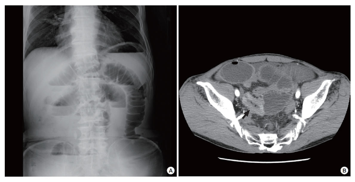
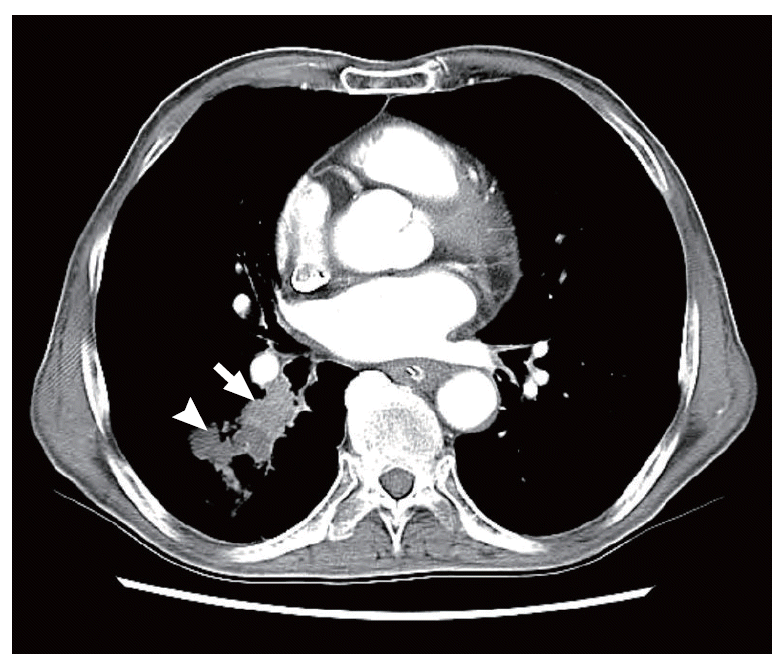
고혈압 외에는 특별한 기저질환이나 과거 수술력이 없었던 73세 남자환자가 2주 전부터 시작된 복통과 복부팽만으로 2차병원에 입원하여 보존적 치료를 받아오던 중 증상의 호전이 없어 본원 응급실로 전원되었다. 내원 당시 신체검사에서 활력지수는 혈압 140/80 mmHg, 심박수 84회/분, 호흡수 18회/분, 체온 36.8°C로 안정적이었으나 혈액검사에서 백혈구는 12,260/μL로 상승되어 있었다. 복부는 심하게 팽만되어 있었고 복부 전체에 압통과 반동압통이 있었으며 장음은 감소된 소견을 보였다. 응급실에서 시행한 복부단순촬영과 복부골반전산화단층촬영에서 회장 원위부에서 조영 증강되는 종괴와 이로 인한 기계적 장폐색의 소견이 보였다(Fig. 1). 동시에 시행된 흉부전산화단층촬영에서는 폐우하엽의 상분절에서 2.3×1.7 cm 크기의 종괴가 발견되었으나(Fig. 2) 기계적 장폐색으로 인한 장손상이 의심되어 이에 대한 정밀검사를 진행하지 못하고 응급개복술을 시행하였다. 수술소견상 회맹부로부터 80 cm 상방의 회장에서 약 4 cm 크기의 종괴가 후복막을 침범한 양상으로 발견되었고 이로 인하여 전반적으로 소장이 늘어나 있었으나 허혈성 변화는 보이지 않아 회장 부분절제술 및 단단 문합술을 시행하였다. 수술 후 병리조직검사에서 회장의 전층을 침범한 중등도분화의 편평상피세포암종으로 확진되었으며 이는 원발성보다는 전이성 소장암으로 강력히 의심되었다(Fig. 3). 수술 후 14일째 폐우하엽의 종괴에 대하여 기관지흡입생검을 시행하였으며 조직검사에서 회장에서 발견된 종괴와 동일한 양상의 편평상피세포암종의 소견을 보였다. 환자에게 원발성 폐암에 대한 추가수술과 항암치료를 권유하였으나 환자는 이를 거부하고 자의 퇴원하였다. 외래추적관찰 3개월째 시행한 복부골반 및 흉부전산화단층촬영에서 각각 다발성 간 전이 및 양측 부신의 전이성 병변이 새롭게 발견되었으며 원발성 폐암의 크기는 4.5× 3.6 cm로 증가된 소견을 보였다. 환자는 수술 후 7개월째 암악액질로 사망하였다.

(A) Plain abdominal X-ray reveals mechanical small bowel obstruction with step-ladder air-fluid pattern in the erect film. (B) Axial abdominal and pelvic computed tomography scan shows abnormal enhancing ileal mass (arrow) with proximal small bowel obstruction.

Axial chest computed tomography scan shows 2.4×1.8 cm soft tissue lesion (arrow) with distal mucus plugging (arrow head) in the superior segmental bronchus of right lower lobe.

The resected segment of the ileum discloses an ulcerofungating/ulceroinfiltrative tumor (A) with pearly white solid cut surface (B) extending to the mesenteric fat. Histologically, the tumor consists show transmurally infiltrating epithelial cells (C) with squamous differentiation (H&E, x100; right inset: H&E, x100; left inset: p63 immunostain, x100).
증례 2
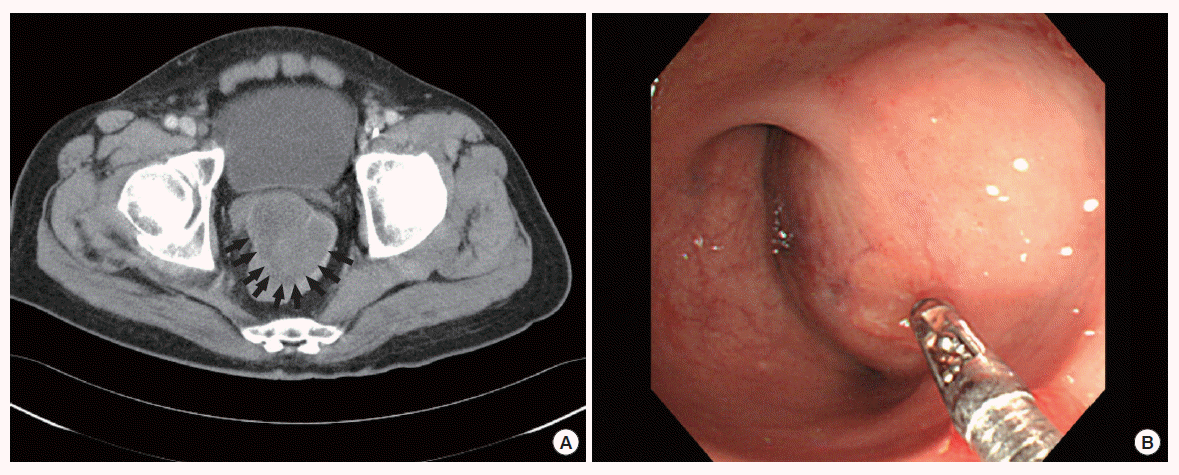
57세 남자환자가 2개월 전부터 시작된 객혈과 우측 전흉부 종괴로 본원 호흡기내과에 내원하였다. 내원 당시 시행한 흉부단순촬영과 흉부전산화단층촬영에서 5 cm 크기의 종괴가 폐 우상엽에서 발견되었다(Fig. 4). 원발성 폐암 및 종양의 흉벽전이 진단 하에 흉부외과에서 폐 우상엽절제술 및 전흉부 종괴절제술을 시행하였고 수술 중 시행된 우측 대흉근의 종괴에 대한 동결절편검사에서 비소세포형 저분화 암종 소견을 보였다. 최종 병리조직검사에서 폐종양은 선편평상피세포암종(adenosquamous carcinoma)으로 확진되었으며 림프절 전이나 신경주위침범은 보이지 않았으나 림프혈관침범 및 벽측흉막침범 소견을 보였다. 환자는 방사선치료를 받던 중 수술 후 3개월 만에 발생된 혈변을 주증상으로 응급실에 내원하였다. 내원 당시 신체검사상 활력지수는 정상소견이었고 복부의 압통이나 반동압통은 없었으나, 혈액검사에서 혈색소가 5.0 g/dL로 심한 빈혈 소견을 보였다. 입원 후 시행한 복부골반전산화단층촬영과 대장내시경상 항문연 8 cm 상방의 직장에서 장경 7 cm 크기의 궤양을 동반한 점막하 종괴가 발견되었으며(Fig. 5) 내시경 조직검사에서 저분화 암종소견을 나타내었다. 환자에게 저위전방절제술을 시행하였으며 병리조직검사에서 종양은 직장의 전층을 침범하였으나 정상점막에서 종양으로 이행되는 부위 없이 점막층을 밀고 올라오는 저분화 암종소견을 보였다. 면역조직화학염색(immunohistochemical staining)에서는 cytokeratin 7과 vimentin에 양성반응을, cytokeratin 20에 음성반응을 나타내었으며 이는 폐종양의 선편평상피세포암종과 동일한 형태로 원발성 폐암의 직장전이로 최종 확진되었다(Fig. 6). 환자는 수술 후 지속적인 우측 대퇴부 통증을 호소하여 시행한 복부골반전산화단층촬영에서 다발성 간 전이와 우측 관골구 및 대퇴골두에 전이성병변이 발견되었으며 저위전방절제술 후 1개월째 사망하였다.

(A) Plain chest X-ray and (B) axial chest computed tomography scans show well-defined 4.8 cm diametered round soft tissue mass with underlying inactive tuberculous lesion in the right upper lobe.

(A) Axial abdominal and pelvic computed tomography scan reveals 6.8×7.0 cm lobulated homogenous low attenuated extraluminal lesion (arrows) in anterior wall of mid-rectum. (B) Colonoscopy shows a huge broad-based submucosal rectal mass, which is located approximately 8 cm proximal to the anal verge.
고 찰
원발성 폐암의 위장관 전이는 대개 점막하 종양의 형태를 나타내는 경우가 많아서 말기까지 증상이 발현되는 경우가 드물며 사후에 부검을 통하여 밝혀지는 경우가 대부분이다[7]. Yoshimoto 등[2]은 IIIB 및 IV기 폐암환자 470예의 부검을 통해서 폐암의 위장관 전이는 11.9%인 56예로 적지 않으며 그 중 소장(8.1%), 위(5.1%), 대장(4.5%) 순으로 전이되었다고 보고하였으나, 실제 임상에서 생존해 있는 환자에게 관찰되는 폐암의 위장관 전이는 여전히 드문 질환으로 알려져 있다. 이는 실제 전이는 존재하지만 증상 자체가 아예 없거나 혹은 있더라도 전반적인 전이에 따른 비특이적인 증상, 또는 항암치료 합병증의 일환으로 유발되는 단순 장염이나 궤양 등에 의한 증상으로 과소평가되기 때문이라고 여겨진다[8]. 따라서 폐암의 위장관 전이에 의해 실제 증상이 나타나는 경우는 0.2%-0.5% 정도로 극히 드문 것으로 알려져 있으며[6,9], 이러한 증상 역시 여타 위장관 질환의 비특이적인 증상과 크게 다르지 않아 진단이 어렵다. 그러나 최근 양전자전산화단층촬영(18F-2-fluoro-deoxy-d-glucose positron emission tomography/computed tomography) 등의 영상의학적 진단기술의 발전과 이를 이용한 원발성 폐암의 병기진단의 관례화, 또한 치료기법의 발전으로 인한 폐암의 생존율 증가로 인해 잠재적인 위장관 전이의 조기진단은 점차 증가될 것으로 생각된다[10]. 일반적으로 폐암의 위장관 전이는 예후가 매우 불량하며 전이의 확진 후 사망까지의 생존기간이 매우 짧아서 Yang 등[1]은 평균생존기간을 130.3일(범위, 23-371일)로 보고하였다. Kim 등[6]은 전이병변의 절제술 후 생존율의 증가를 주장하기도 하였으나, 진단 당시 전이의 정도가 심하거나 천공이나 출혈 등의 합병증이 동반된 경우 예후가 불량하다는 것이 보편적이다[1,11]. 이는 주로 위장관 전이가 폐암의 말기에 발생되고 대부분 위장관 이외의 타 장기에도 전이가 동시성 또는 이시성으로 발생한다는 사실에 기인하는 것으로 생각된다. 본 증례의 경우도 크게 다르지 않아서 회장전이의 경우는 수술 후 3개월 만에 간과 부신에 전이가 새롭게 발견되었고 7개월만에 사망하였으며, 직장전이의 경우는 수술 후 불과 15일만에 간과 대퇴골에 전이가 발견되었으며 1개월만에 사망하였다.
폐암의 소장전이는 가장 흔한 위장관 전이로 알려져 있다. 발병기전은 혈행성과 림프성 전파를 통해 파급된 종양세포가 점막하층이나 근층에 초기 병소를 형성하고 크기가 커진 후 장벽의 전부 또는 일부를 침범하여 결과적으로 궤양출혈 및 천공 또는 장폐색을 유발하는 것으로 알려져 있다[1]. McNeill 등[11]은 소장전이가 있는 15명의 환자들 중 장폐색은 1명에서만 발생되었고 14명에서 천공이 발생되어 소장천공이 가장 흔한 임상양상이라고 보고하였으며 이는 폐암에 의한 소장전이가 다른 전이성 암보다 장폐색을 유발할 정도로 병변이 커지기 전에 괴사를 조기에 일으켜 천공이 상대적으로 흔하다고 설명하였다.
폐암의 직장전이는 의학 분야 데이터베이스인 PubMed와 Embase에서 lung cancer, rectal metastasis, gastrointestinal metastasis 등의 색인단어를 입력하여 검색한 결과 문헌상 현재까지 전세계적으로 단 3예 만이 보고되어 있을 정도로 극히 드물다. Johnson과 Allen [3]이 1995년 소세포폐암의 이시성 직장전이로 복회음절제술 후 19개월 만에 사망한 증례를 세계 최초로 보고하였고 Cedres 등[4]은 수술적 치료 없이 폐생검과 직장생검을 통해 확진된 비소세포폐암의 이시성 직장전이를 보고하였으며 Miyazu and Kobayashi [5]은 원발성폐암 수술 후 부신과 직장에 발생한 다발성 이시성 전이를 보고한 바 있다. Johnson과 Allen [3], Cedres 등[4]이 보고한 증례는 각각 직장출혈과 복부통증에 의해 발견된 증상이 동반된 직장전이였으나 Miyazu 등[5]은 폐암치료과정 중 시행된 양전자단층촬영에서 우연히 발견된 직장전이였다. 본 증례 중 두 번째 증례 역시 혈변을 동반한 폐암의 직장전이로서 한국의학논문데이터베이스(kmbase.medric.or.kr)를 통해 PubMed, Embase에 등재되어 있지 않은 국내 논문까지 검색한 결과 국내 최초로 보고되는 것으로 생각한다.
원발성 폐암의 위장관 전이는 일반적으로 폐암 진단 후 이시성으로 발견되는 경우가 대부분이나 본 증례와 같이 장출혈이나 장폐색 또는 장천공 등의 심각한 합병증을 동반한 동시성 전이로 발견되는 경우 역시 적지 않아서 응급개복술을 필요로 하기도 한다. 따라서 폐암환자에서 비특이적인 위장관 증상이 동반된 경우 일차적으로 소화성 궤양, 단순장염 등의 급성복증을 의심해야 하지만 위장관 전이도 고려해야 하며, 복막염 등으로 응급개복술을 시행 후 위장관의 전이암을 발견하여 원발암을 찾아야 할 경우라면 폐암에 대한 정밀검사도 고려해야 할 것으로 생각된다.
Notes
No potential conflict of interest relevant to this article was reported.
Acknowledgements
This work was supported by INHA University Research Grant.
