대장 장간막에 발생한 원발성 신경 내분비 종양
Primary neuroendocrine tumor of the colon mesentery
Article information
Trans Abstract
Primary neuroendocrine tumor of the mesentery is very rare. More than 90% of gastrointestinal neuroendocrine tumors are located in the appendix, small intestine and rectum. We present a case of very rare primary neuroendocrine tumor of the mesentery showing malignant character though large size. The patient visited Yeungnam University Medical Center presented with palpable abdominal mass which had known occasionally. At laparotomy, a huge mass was found to be arising from transverse colon mesentery and mass was invasion to transverse colon and anterior antrum of the stomach. The mass was measured as 20.5×15.0 cm sized that demonstrated a malignant character. At 19 months of follow-up, the patient was recurrence free.
INTRODUCTION
Most of the neuroendocrine tumors (NET) of the gastrointestinal tract are traditionally termed “carcinoid tumors.” More than 90% of gastrointestinal carcinoids are located in the appendix, small intestine and rectum [1]. Carcinoid tumor is the most frequent primary malignant tumor of the small intestine beyond the ligament of Treitz and most frequently arises in the terminal ileum [2,3]. Secondary mesenteric involvement of small bowel carcinoid tumors is common, reported as occurring in 40% to 80% of cases [3]. However, primary mesenteric carcinoid tumors are very rare and most solid tumors arising in the mesentery are usually metastatic tumors [2]. The management of patients with metastatic NETs is focused on the palliation of symptoms and on the prevention of further complications of the disease. We present a very rare case of a primary mesenteric neuroendocrine tumor with 20.5×15.0 cm sized that demonstrated a malignant character.
CASE REPORT
A 72-year-old man presented with palpable abdominal mass which had known occasionally. The patient had abdominal obesity. So he didn’t know intra-abdominal palpable mass. Physical examination revealed an huge round and fixed mass with mild tenderness in the periumbilical area. Computed tomography (CT) scan demonstrated an 15×10 cm sized well-encapsulated malignant appearing mass with located in the transverse colon mesentery and it attached to great curvature site of stomach (Fig. 1). Barium enema demonstrated more than 10 cm of mesenteric border of the transverse colon was blurred with mass but the impairment of contrast passage or peristaltic movement was not seen (Fig. 2). At laparotomy, a huge mass was found to be arising from transverse colon mesentery and mass was invasion to transverse colon and anterior antrum of the stomach (Fig. 3). Some amount of ascitic fluids were presented and we sent to analyze the ascitic fluids at pathologic department. We carefully dissected and removed the mass. We did transverse colectomy and wedge resection of the stomach.

Computed tomography scan shows huge well-defined mass in colon mesentery and attached to great curvature site of stomach
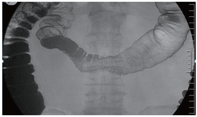
Barium enema shows more than 10 cm of mesenteric border of the transverse colon was blurred with mass but the impairment of contrast passage or peristaltic movement was not seen
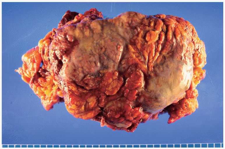
Well-capsulated mass originated transverse colon mesentery.
The specimen was well-encapsulated and surface showed heterogenous fibrous appearance. Hemorrhage and mucinous material was identified. Histological examination showed tumor cells were malignant small cells and tumor necrosis was present (Fig. 4). Immunohistochemistry showed the tumor cells were positive for neuroendocrine markers (synaptophysin and CD56) (Fig. 5) and cytokeratin, CD99, chromogranin A were also positive. Ki-67 was approximately 70% positive. Surgical pathologic report showed carcinoma with neuroendocrine differentiation, consistent with neuroendocrine carcinoma, high grade. Tumor size was 20.5× 15.0× 9.8 cm and tumor necrosis was present. Regional lymph node were no tumor metastasis (0/5). Cytology of ascitic fluids was reported to class V (cytology conclusive of malignancy). At postoperative 19 months of follow-up, the patient was asymptomatic and recurrence free.
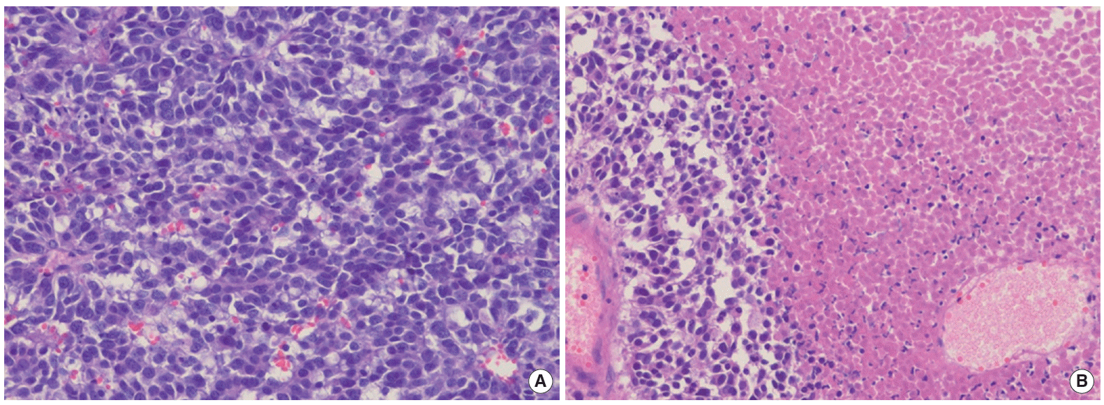
(A) Histological examination showed tumor cells were malignant small cells and (B) tumor necrosis was present. H&E, ×200
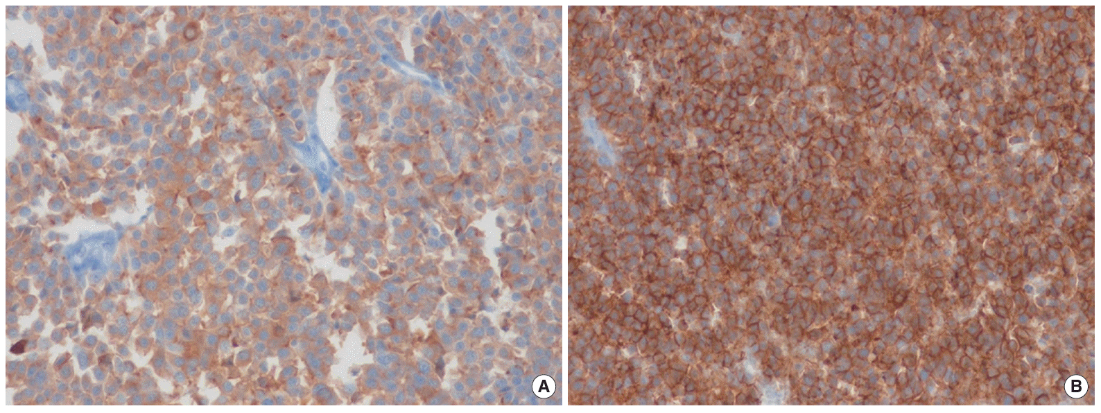
Tumor expresses strong positivity in immunohistochemical stain with synaptophysin (A) and CD56 (B). ×200
DISCUSSION
Carcinoid tumor is a rare, slow-growing, NET with about 90% of the lesions arising in the gastro intestinal track (GIT) [1]. GIT carcinoid tumors are classified by the embryologic origin as foregut, midgut and hindgut. 46% to 64% of GIT carcinoid tumors arise in the midgut and most midgut carcinoid tumors originate in the terminal ileum [3]. However, primary carcinoid tumors of the mesentery are very rare [2].
On CT scan, mesenteric carcinoid tumors exhibit varying degrees of fibrosis, calcification, focal or diffuse neurovascular bundle invasion by the tumor or both mechanisms [2]. Surgical excision is a mainstay of treatment for carcinoid tumor. Larger tumors are usually associated with locally advanced or distant metastasis [3]. The morphological and immunohistochemical features place the described tumor in the NET group. However, the subclassification of these tumors is not as well defined as in the lung. The tumor’s large size and extensive necrosis are adverse features supported by the presence of a metastatic lymph node. However, the low mitotic rate and low proliferation rate (rather sparse Ki-67 staining) and the only mild to moderate cytologic pleomorphism do not enable the diagnosis of a high grade NET. Therefore, the diagnosis of an intermediate grade NET (or atypical carcinoid) seems to be the most appropriate diagnosis [4]. NETs have specific immunohistochemistry features. Synaptophysin, chromogranin A, cytokeratins and neuron- specific enolase are usually positive [1]. In our case, synaptophysin, chromogranin A and cytokeratin were positive, which confirmed the diagnosis of NET. To make the diagnosis of mesenteric NET, one must first rule out other primary sites by the use of CT, colonoscopy, small bowel series and scintigraphy. Abdominal CT and an octreotide scan exhibited a mesenteric tumor and liver metastases. A colonoscopy, small bowel series and surgical exploration of the abdomen can confirm the diagnosis of mesenteric NET [5].
In this case, the tumor originated from the transverse colon mesentery and directed invasion to the stomach. But it was well encapsulated and free from the small intestine. At the time of surgery, whole abdominal cavity especially the entire small bowel was meticulously inspected and no evidence of tumor mass or enlarged lymph node was found. There was no evidence of tumor anywhere else in the abdomen including the liver and other solid organs. So, it may be a primary mesenteric NET. Our pathologic biopsy reported tumor cells were malignant small cells and positive for synaptophysin, CD56, cytokeratin, CD99, and chromogranin A. Ki-67 was approximately 70% positive. Surgical report showed carcinoma with neuroendocrine differentiation, consistent with neuroendocrine carcinoma, high grade. We studied CT scan and barium enema. At laparotomy, we carefully dissected the mass and did transverse colectomy and wedge resection of the stomach. This case shows a rare large primary mesenteric NET with characteristics of uncommon position with malignant nature and has not been cited in the current literature. This patient has several poor prognostic factors such as tumor necrosis, presence of cytokeratin, cytology class V and old age. But postoperative 19 months of follow-up, the patient was asymptomatic and recurrence free.
Notes
No potential conflict of interest relevant to this article was reported.